What is ultrasound and how does it currently work?
Ultrasound, or sonography, uses sound waves to map structures inside the human body. Although pregnancy comes to mind when talking about ultrasound, ultrasound has a variety of other uses such as diagnosing diseases, evaluating blood flow, guiding the needle during a biopsy, checking the thyroid gland, and observing joint inflammation.Humans can hear sound frequencies ranging from 20 Hertz (Hz) to 20000Hz, but ultrasound is imperceptible to humans because it has a frequency above 20000 Hz. Ultrasound can be compared to sonar, the imaging method used by submarines to detect missiles or other ships around them.
Most ultrasound systems employ the use of a transducer, which is composed of an array of piezoelectric crystals. Transducers typically convert one form of energy into another, and the transducers used in ultrasound systems are no different. When electricity is applied an ultrasound transducer, it emits high frequency sound waves that bounce off of the variety of tissues in the human body. Based on the tissues' properties (density, smoothness, hardness), the waves are reflected with different frequencies and wavelengths. When these reflected waves reach the transducer again, another electrical current is generated, allowing the ultrasound to display a point.![]()
While transducers are usually placed above the body, there are instances in which it must be placed inside an opening:
- Transesophageal echocardiograms requires the transducer to be inserted into the esophagus (throat) to get clear images of the heart.
- Transrectal ultrasound is when the transducer is placed into the rectum to get images of the prostate.
- Transvaginal ultrasounds are when a transducer is inserted into the vagina to observe the uterus and ovaries.
Once an ultrasound is complete, radiologists analyze the images for any signs of disease, helping cure or even prevent diseases.
So what is a wearable ultrasound?
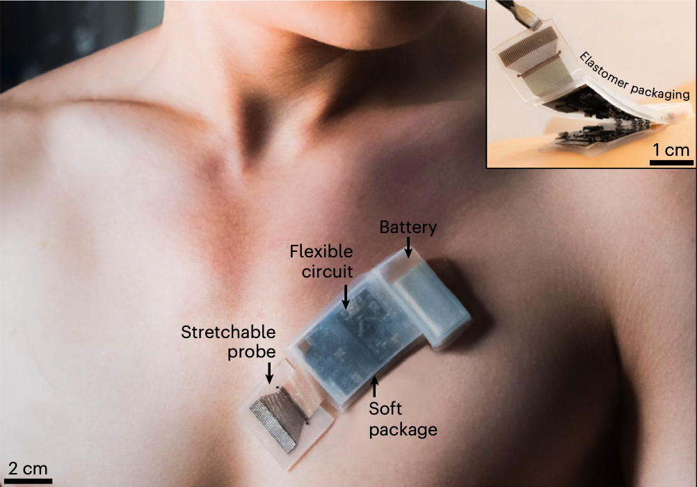
Recently, scientists at the University of California, San Diego have created a wireless wearable ultrasound device that can create images of internal tissues while the patient moves around freely. While traditional ultrasounds require that the patient remain still and close to the handheld, wired device, this new wearable sticks to the body and can take useful measurements while patients exercise or conduct other activities.