Biomedical Devices in the News
You may have recently seen some articles about how a man paralyzed from the waist down is now able to walk with the help of a brain-spine interface. A great example of how biomedical devices have the potential to improve the lives of people with disabilities, brain-spine interfaces are a relatively new technology that have a wide variety of uses within the medical world.What is a brain-spine interface?
To understand how brain-spine interfaces work, we first have to understand how the brain sends signals to the rest of the body.The brain is composed of billions of neurons. When the brain attempts to call on information to achieve a purpose, neurons in different parts of the brain 'fire'. They send out an electrical signal that travels from neuron to neuron, through nerves, and finally to the selected organs. When moving muscles, a certain type of neuron - motor neurons - are triggered.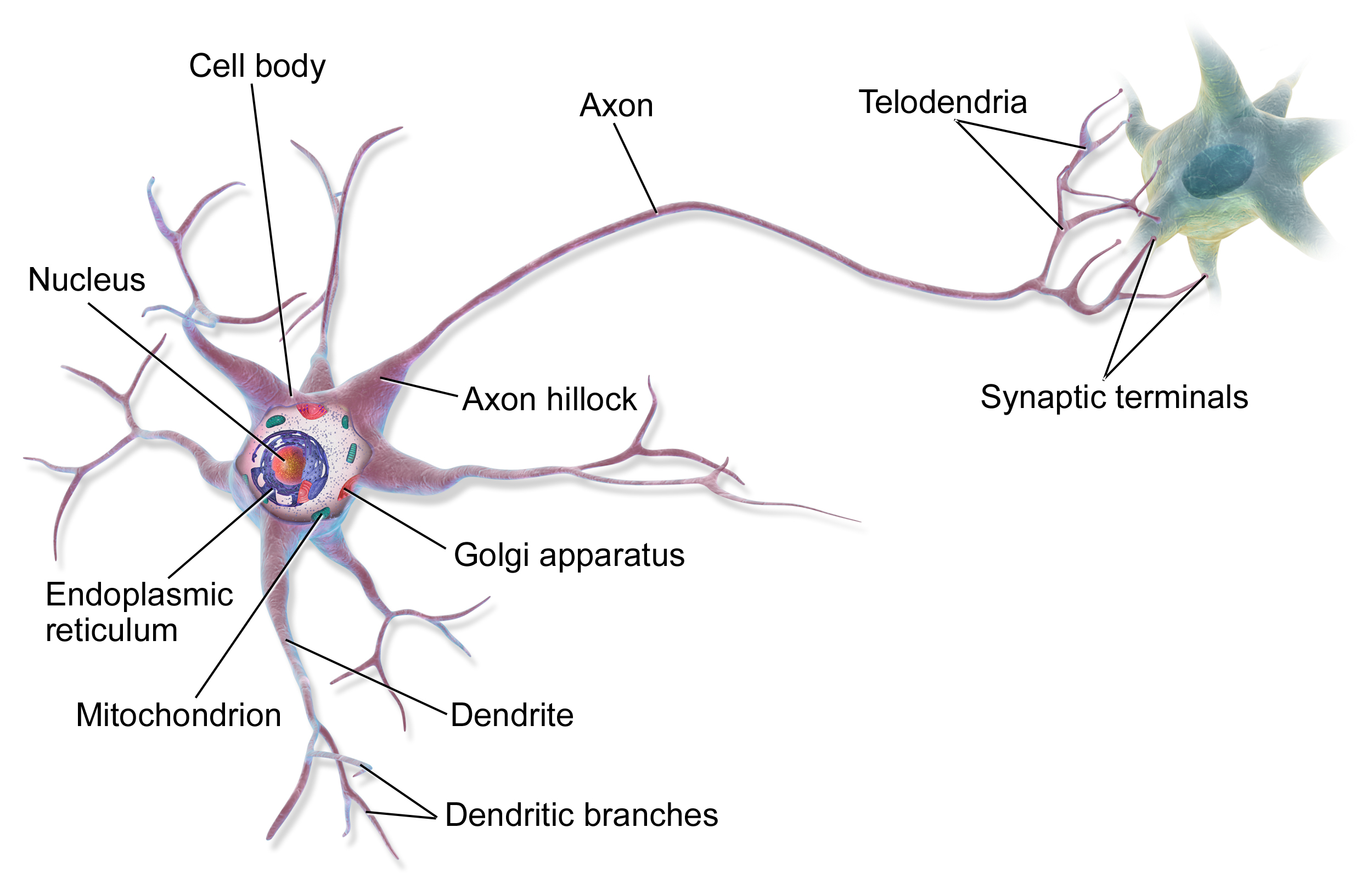
For the patient in these news articles, the only injury was to the neck. Instructions sent from the brain could not pass through nerves in the neck, so they never reached the spinal cord. Thus, a brain-spine interface is a digital bridge that externally connects the brain and spinal cord, bypassing nerves and neurons located in the nervous system.
Recording Signals from the Brain
The first part of establishing a working brain-spine interface is recording signals from the brain. Different parts of the brain are responsible for different groups of actions. Motor movement is handled by the sensorimotor cortex.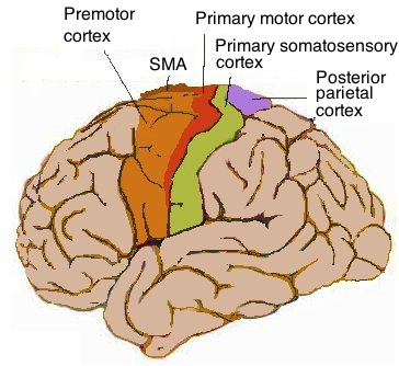
Transmitting and Delivering the Signals
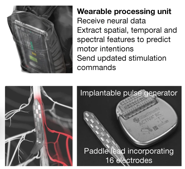
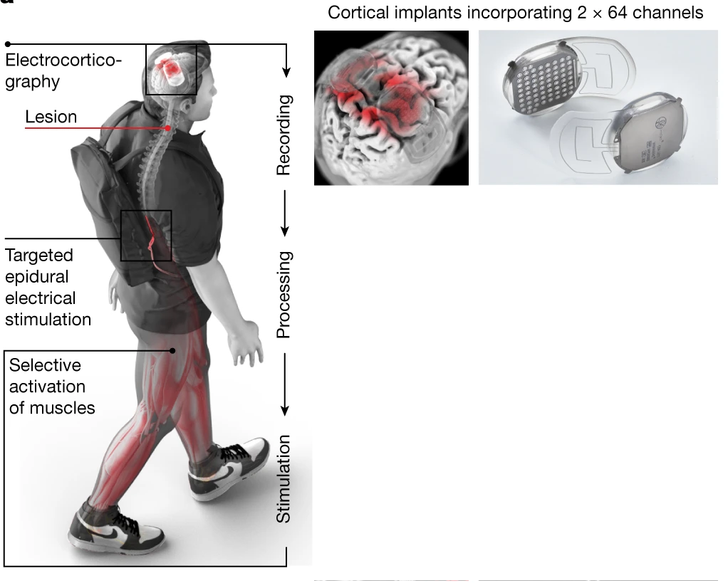
Now that the signals have been recorded, they must be sent to a processing unit for analysis. After the unit determines which part of the lumbosacral spinal cord must be activated, it must send the signal to a device that can induce neuron activation.The wearable processing unit is located inside the patient's backpack. To prevent the patient from being bogged down by wires connected to the brain, the cortical implants wirelessly deliver information to the processing unit. After extracting the spatial, temporal, and spectral features of the signals, it calculates the best location along the spinal cord to be stimulated (each segment of the spinal cord triggers a different muscle to be activated). This location is wirelessly transmitted to a paddle lead, an electrical device that is surgically implanted into the spinal cord. Consisting of 16 electrodes, the paddle creates an electrical signal that activates the spinal cord, leading to the activation of leg muscles. Finally, the patient has regained their ability to walk naturally.