What is cell division?
Cell division is the process by which a cell reproduces or creates a copy of itself. There are two main phases in cell division: nuclear division and cytokinesis. Nuclear division is when the nucleus divides and cytokinesis is when the cell's cytoplasm divides. Nuclear division also consists of two different methods: mitosis and meiosis. Mitosis results in cells that have the same genetic material as the original cell, while meiosis results in cells with half the genetic material of the original cell.
Mitosis and meiosis begin with the genetic material in the form of chromatin condensing into tightly coiled chromosomes. Chromosomes are composed of two sister chromatids that are joined at the centromere. Each sister chromatid consists of a tightly coiled DNA molecule.
Diploid cells consist of two copies of each chromosome, with one from the paternal parent and the other from the maternal parent. Each diploid cell contains 46 chromosomes (or 23 homologous pairs of chromosomes) if there are no problems in the cell.
The cell cycle regulates the life of the cell and whether or not the cell divides. The first part of the cell cycle before mitosis or meiosis occurs is called interphase. In this phase, the cell prepares for mitosis or meiosis during three stages: G0, S, and G2. During interphase, the cell's chromosomes condense, the cell's genetic material condenses, and microtubule organizing centers (MTOCs) are created. MTOCs play a major role during mitosis and meiosis.
During G1 phase, the cell grows and starts to get ready for the cells chromosomes to divide.
In S phase, the cell's chromosomes and MTOCs duplicate.
In G2 phase, the cell ensures that it is ready to start mitosis/meiosis.
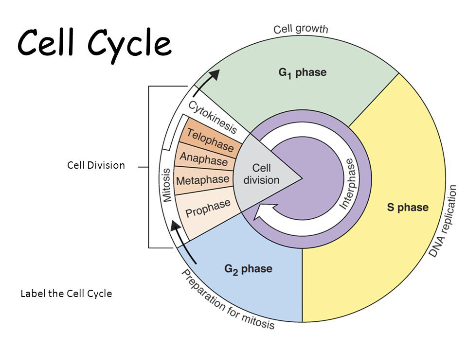
Mitosis
Mitosis is composed of four stages:
- Prophase is when the cell's chromosomes condense, the nucleoli disappear, and the nuclear envelope disintegrates. A mitotic spindle forms from the MTOCs, which move to opposite poles of the nucleus. As the microtubules move toward each other, they join to a region in the centromeres of the chromosomes, called kinetochores.
- Metaphase is when the chromosomes align at the metaphase plate, which is usually near the center of the cell's nucleus.
- Anaphase is when the microtubule spindles move apart, and in the process, pull the sister chromatids of each chromosome to opposite poles of the cell. The microtubules are shortened because tubulin units that make up the microtubules are removed. Since each pair of sister chromatids is separated into two sister chromosomes, each daughter cell will have the same number of chromosomes as the original parent cell.
- Telophase is when the cell returns to its normal state (before it underwent division). The nuclear envelope reforms and two nuclei are formed. Each nuclei contains all the chromosomes at one of the two poles. Additionally, the nucleoli reappear. The final step is cytokinesis.
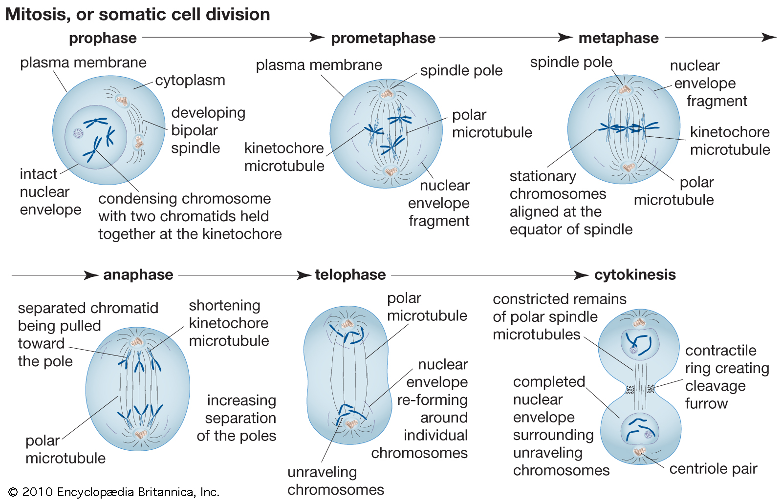
Cytokinesis
Cytokinesis is the final step of mitosis/meiosis. The cell's cytoplasm divides and two new cells are formed. Cytokinesis depends on the cell type.
A cell plate is formed in plant cells, and a cleavage furrow is formed in animal cells. The cell plate is composed of a number of vesicles that enlarge and eventually fuse to form the plate. The cleavage furrow is formed as actin filaments make a ring around the animal cell's plasma membrane. As the ring narrows and closes, the cell is divided into two daughter cells.
Meiosis
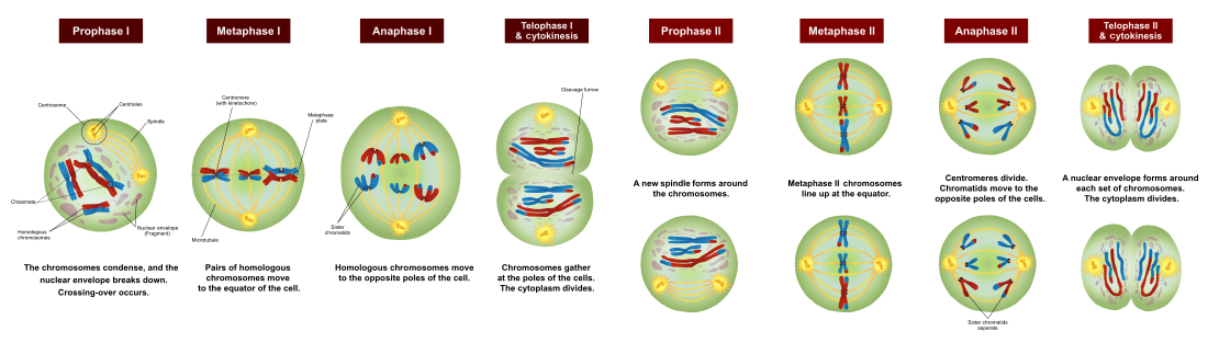
Meiosis is similar to mitosis, but its primary purpose is to crete gametes. Unlike mitosis, meiosis consists of two sequences of divisions: meiosis I and meiosis II. Following are each of the phases in meiosis:
- Prophase I is the stage when the nucleolus disappears, the nuclear envelope disintegrates, and the spindle forms. Unlike prophase of mitosis, recombination between pairs of homologous chromosomes occurs. During a process called synapsis, chiasmata form and the chromosomes exchange genetic material.
- Metaphase I is when homologous pairs of chromosomes align at the metaphase plate. Spindle fibers attach to the pairs of homologous chromosomes.
- Anaphase I is when the spindle fibers separate the pairs of homologous chromosomes and move them ot opposite poles of the cell.
- Telophase I is the stage where the nucleus reforms and two cells with the same number of chromosomes as the original parent cell are formed. NO REPLICATION OCCURS BEFORE PROPHASE II.
- Prophase II is when the nuclear envelope disappears again and a spindle is formed again. There is no crossing over.
- In metaphase II, the chromosomes align on the metaphase plate and the spindle fibers attach to each chromosome.
- Anaphase II is when the separation of the chromosomes occurs, and the sister chromatids move to opposite sides of the cell.
- In telophase II, the nuclear envelope is formed again. There are now 4 cells and each one has half the number of chromosomes in the original parent cell.
Main differences between mitosis and meiosis.
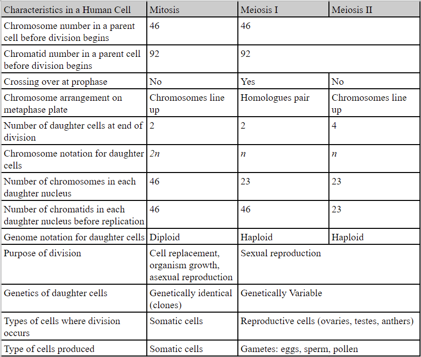
Mitosis only occurs in somatic cells, while meiosis only occurs in reproductive cells and produce gametes. Eventually, the gametes combine and in a process called fertilization, a diploid zygote forms. The zygote repeatedly divides and gives rise to a new multicellular organism.
Genetic variation
Look around and you'll find that all humans are not identical and everyone is a little bit different. But what are the reasons for this difference? Genetic recombination allows for reassortment of genes and unique organisms. There are 3 main processes that contribute to genetic recombination:
- Crossing over during prophase I of meiosis leads to a random exchange of DNA between non-sister chromosomes.
- The independent assortment of homologues during metaphase I of meiosis leads to a random arrangement of chromosomes at each pole of the cell.
- The random joining of gametes also leads to a different combination of genetic material in the zygote (because each sperm and egg contains a different set of genes).
Cell Cycle Regulation
The cell cycle is regulated by a number of factors. If the cell cycle is unregulated, the cell can turn into a cancer cell.
The surface area to volume ratio of the cell limits the cell's size. Since the volume of the cell grows faster than the surface area of a cell, the SA/V ratio decreases as the cell grows. A large volume compared to the cell's surface area makes it harder for the cell to allow substances to diffuse through the cell. To remedy this problem, the cell must divide.
The genome to volume ratio must also be controlled. The genome consists of all the genetic material that is stored in chromosomes. If the volume of the cell is too big, the genome will be unable to produce enough materials to control cellular activities.
The cell cycle is also regulated by checkpoints:
- The G1 checkpoint is near the end of the G1 phase. If DNA damage is detected, the cell performs apoptosis. If the cell does not need to divide, it remains in the G0 phase.
- The G2 checkpoint is at the end of the G2 phase. Just like the G1 checkpoint, if DNA damage is detected, apoptosis occurs.
- The M checkpoint is during metaphase and it ensures that all the microtubules are connected to the kinetochores at the metaphase plate.
Additionally, cyclins and cyclin-dependent kinases (Cdks) ensure that the cell either advances or stops at the checkpoints.